make preferable
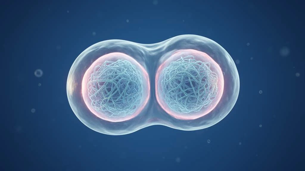
In the earliest stages of embryonic development, not only genetic information but also the physical state of the cell in which it is stored plays an important role. Scientists have discovered that shortly after fertilization, the cell nuclei of mouse embryos briefly become much softer and more pliable. This enables the activation of genes that initiate the developmental program of the future organism. The discovery reveals a previously unknown mechanism of early embryonic development and could help researchers investigate why some embryos fail to develop normally.
The study was published in the journal Nature. An international team of scientists examined how the shape and mechanical properties of cell nuclei change in the first hours after fertilization.
The nucleus temporarily loses its stiffness
After fertilization, the egg begins to divide. In its early development, the embryo passes through successive stages: first it consists of one cell, then two, four, and increasing numbers of cells. During this time, a crucial transition occurs: the embryo gradually begins to control the activity of its own genome.
At the two-cell stage, the so-called zygotic genome is activated—a set of genes that becomes active after fertilization and supports further development. Until this point, RNA molecules and proteins accumulated in the egg before fertilization play the main role in controlling early processes.
The scientists found that cell nuclei acquire unusual mechanical properties precisely during the activation of the zygotic genome. Unlike nuclei at the one-cell and four-cell stages, which remain predominantly rounded, the nuclei of a two-cell embryo gradually become deformed. These changes begin approximately 21 hours after fertilization, peak at 24–26 hours, and gradually disappear by 32–36 hours.
To measure not only the shape but also the stiffness of the nuclei, the researchers developed a specialized system using a nanocapillary. Its fine tip gripped the surface of the nucleus and gently stretched it, allowing the researchers to assess its resistance to deformation.
The results showed that nuclei become significantly softer at the two-cell stage. Their stiffness fell from approximately 5 nanonewtons per micrometer at the one-cell stage to 2 nanonewtons per micrometer. At the next stage, when the embryo consisted of four cells, the value rose again to approximately 4.6 nanonewtons per micrometer.
This is therefore not a random change in shape, but a brief transition to a specific physical state that the embryo needs at a particular point in its development.
The protein that makes the nucleus flexible
The researchers also identified the molecular mechanism responsible for the temporary softening of the nucleus. A key role is played by lamin B1, a protein that forms part of the nuclear envelope. Lamins form a supporting structure that helps the nucleus maintain its shape and mechanical strength.
At the two-cell stage, the amount of lamin B1 at the nuclear envelope drops sharply—by approximately 70%. This process is linked to autophagy, a mechanism by which the cell breaks down and recycles its own components. In this case, the protein LC3 helps remove lamin B1 by directing it for degradation through the autophagy–lysosome system.
When the researchers prevented LC3 from interacting with lamin B1, the protein remained at the nuclear envelope, and the expected softening did not occur. The development of these embryos was impaired. This established a link between lamin B1 removal, changes in the mechanical properties of the nucleus, and the normal activation of the genetic program.
But why is nuclear flexibility so important for gene activation?
Inside the nucleus, DNA is organized together with proteins into chromatin. For the necessary genes to become active, the cell must provide access to specific regions of the genome and activate the processes that read genetic information. The scientists found that in soft nuclei, active RNA polymerase II—an enzyme involved in synthesizing RNA from a DNA template—accumulates near the nuclear envelope. Some key regions of the genome responsible for initiating development are also located there.
Additional experiments showed that artificially increasing nuclear stiffness interferes with these processes: transcriptional activity decreases, and the expression of important genes involved in zygotic genome activation is suppressed.
Could this help assess embryo quality?
The scientists tested whether nuclear stiffness was associated with an embryo’s subsequent ability to develop. To do so, they monitored embryos after taking measurements at the two-cell stage.
All 66 embryos with soft nuclei included in the corresponding experimental group reached the blastocyst stage on schedule—an early developmental stage at which the structure needed for subsequent implantation in the uterus forms. Among 15 embryos with impaired development, seven were found to have increased nuclear stiffness.
A similar characteristic was also identified in embryos produced by somatic cell nuclear transfer, a technique used, among other applications, in cloning.
The study’s authors believe that the mechanical properties of the nucleus could serve as one indicator of normal early development. However, the findings so far come from mouse embryos. Further research is needed to determine whether this mechanism also applies to humans and whether it can be used to assess embryos in reproductive medicine.